Complications of Spinal Cord Injury
Complications of Spinal Cord Injury

Having a spinal cord injury is not just an isolated medical event. People with SCI often suffer from secondary complications of having the disorder. Some of the complications are mere nuisances, while others can be life-threatening.
If you or a loved one has sustained a spinal cord injury in an accident caused by someone else, you may be eligible to pursue compensation for your damages and losses. Our injury lawyers can help guide you through the legal process of filing a personal injury claim. Call us at (916) 921-6400 for free, friendly advice.
In this article:
- Autonomic Dysreflexia
- Deep Vein Thrombosis
- Spasticity
- Heterotopic Ossification
- Pressure Sores
- Pain
- Syringomyelia
This is a medical emergency associated with SCI patients who have lesions at T6 or higher. A few patients with lesions as low as T8 can suffer from the condition. It can result in extreme headache, high blood pressure, and an increased risk of getting strokes or seizures. People with SCI have died from autonomic dysreflexia. Most people have their first episode under the direct and competent care of healthcare professionals while they are in rehab; a select few won’t have an episode until they are home, which can result in a trip to the emergency department.
Autonomic dysreflexia usually happens in the context of some sort of bodily insult that would normally result in pain if the patient had normal sensation. People with SCI do not have the sensory capabilities to send signals to the brain in order to remedy the cause of the pain so the autonomic nervous system goes into hyper-drive, leading to blood pressure elevation and heart rate lowering. The face becomes flushed and things like nausea, headache, anxiety, nasal congestion, and irritability are common. Vision changes can occur because of extremely high blood pressure. Sweating of the upper half of the body is common as well, with coldness to the skin below the level of the lesion.
The treatment of autonomic dysreflexia is to sit up so that blood can pool around the feet as well as finding the source of the symptoms and correcting the underlying problems. The most common cause of AD is having a bladder that is much too full. It can occur when self-catheterization is not done in time or when an indwelling catheter becomes blocked so that urine doesn’t drain into the bag but fills up the bladder. Even catheterizing someone can trigger the symptoms of AD. This is why anesthetic jelly is recommended for catheterization even though the patient cannot technically feel any pain from the catheter.
Things like bladder infections, pressure sores, constipation, ingrown toenails, burns, and tight clothing can trigger autonomic dysreflexia. If the cause of the symptoms is not readily noticeable, emergency medical help should be summoned. The main treatment of AD in which the underlying cause cannot be found or eliminated includes blood pressure lowering medications.
The best way around autonomic dysreflexia is to prevent it from occurring in the first place. Catheterization should be regularly scheduled and a bowel program should be adhered to. Frequent checks for potentially painful conditions like pressure sores and ingrown nails should occur so as to prevent the AD symptoms from occurring.
SpasticityMany people with spinal cord injuries develop involuntary increases in muscle tone—a condition called spasticity. The muscles are very stiff and it is more difficult to move the affected extremities because of the stiffness. Spasms of the muscles can occur periodically. Spasticity comes from a spinal cord reflex that occurs when the brain fails to send signals to the muscles to stop contracting. Spasticity can involve intermittent spasms only or it can lead to chronic stiffening of the muscles. There can be flexion spasms or extension spasms, particularly of the lower extremities, which can be quite painful. Upper extremity spasticity can result in a completely clenched fist that does not open well for handwashing.
Spasticity doesn’t have to be a bad thing. Paralyzed patients can use their spasticity to control trunk posture and can get in and out of bed easier because the muscles aren’t flaccid and “dead weight”. Spasticity of the lower extremities might also prevent deep vein thrombosis of the lower extremities.
Spasticity increases may signal different things, such as a urinary tract infection, pressure sore, kidney stones, oncoming autonomic dysreflexia, and ingrown toenails. In that way, an increase in spasticity can be warning signs that something needs to be looked into.
Mild spasticity can be treated with stretching exercises or modified yoga. Some people will wear splints to keep the extremities in the proper alignment. Medications to control spasticity are given to patients who still have spasticity after natural measures have failed. If just one area is affected, neurolysis can be performed in which medication is injected directly into the affected nerve. Alcohol and phenol have been used for this purpose. The alcohol and phenol blocks are generally permanent and are done by a physiatrist or other skilled physician. Botulinum toxin has become the agent of use in recent years for muscle spasticity. Repeat injections must be done every 3 months or so. A baclofen pump can be inserted surgically, which provide anti-spasticity effectiveness at much lower doses so that side effects can be minimized. The pump is refilled by the doctor every month or two.
Deep Vein ThrombosisDeep vein thrombosis is another side effect of spinal cord injury. This is due to their relatively slow blood flow through the peripheral veins. The blood can clot in the distal veins, leading to deep vein thrombosis. In severe cases, a DVT can break off of the main clot and can travel to the lungs, causing a potentially life-threatening condition called a pulmonary embolism. Elastic compression bandages can be used to keep blood flowing through the veins and to prevent stagnation of blood. Some people need to be on a blood thinner to prevent blood clots from forming.
Watch the video below for more on deep vein thrombosis.
Heterotopic OssificationThis is when small areas of bone crop up in non-bony areas, usually near the joints. No one knows why these deposits develop in SCI patients. The heterotopic bone is usually only in areas of the body affected by paralysis. The signs of heterotopic ossification include redness, swelling, and pain around the joint or joints affected by the disease. Joint mobility can become limited. One test of heterotopic bone formation is the alkaline phosphatase blood test, which is elevated when the condition is occurring. Radioactive tracer has been used to identify areas of new bone formation even when it doesn’t yet show up on X-ray. The treatment is to remove the areas of ossification and to practice passive range of motion exercises. The drug, etidronate, has been found helpful in the management of the inflammation associated with the condition.
Pressure SoresPressure sores are caused by sitting or lying too long in one position. When being in the same position for too long a period of time, ischemia forms in that area and the skin breaks down. Common areas in wheelchair-bound individuals are the hips and thighs. Pressure sores can become so deep that they affect muscle and bony tissue. Infection is a secondary complication and these can be very difficult to treat. Most SCI patients will develop a pressure sore at some point in their lives. The best treatment for bed sores is to prevent them by doing meticulous skincare and changing positions at regular intervals. Skin can also break down because of ill-fitting braces and from wheelchair cushions. Shifting may need to be done as often as every fifteen minutes. Some paraplegics wear watches that go off every fifteen minutes to remind them to change positions.
Many SCI patients sleep on foam, air beds or gel beds to reduce pressure sores during the night. Splints for the heels can reduce pressure sores in the heel area. Some of the best treatments for the prevention of bedsores include having good nutrition with adequate protein and vitamin content in the meals. Protein intake is increased when pressure sores occur so that the sores can better heal. Smoking and alcohol cannot be taken by SCI patients as these contribute to pressure sore formation.
The only way to heal a pressure sore is to debride the dead tissue away, allowing the remaining healthy tissue to grow and heal the wound. Wet to dry dressings or surgical debridement can be used to heal the wounded area. Whirlpools and enzymatic treatments can also debride wounds. Electrical stimulation to the pressure sore area has been found also to maximize the healing potential of these types of wounds. Vacuum-assisted closure will help heal difficult wounds. Some people will need skin grafting and/or flap repairs to close the affected areas.
PainPain is experienced by most people with SCI. This pain is poorly understood and is difficult even for the patient to describe what kind and at what location the pain is. Musculoskeletal pain happens particularly around joints, muscles, bone, and connective tissue. Overuse of the upper extremities in paraplegics can lead to upper extremity pain. Rest and nonsteroidal anti-inflammatory pain medications are the treatment of choice for musculoskeletal pain. Carpal tunnel pain can happen because of repetitive motion to the affected arms and this is treated with splints and/or surgery to open up the carpal tunnel in the wrist.
Neuropathic pain comes from damaged nerves in the brain and/or spinal cord. Peripheral nerves can also elicit pain. People with this type of pain describe ‘pins and needles’, burning pain, or an electric-shock type of pain in various areas. This type of pain gradually diminishes over the years. This type of pain is similar to the phantom pain exhibited by amputees.
Visceral pain is caused by pain in internal organs or soft tissue structures. It can happen with fecal impactions, bowel perforation, or bowel infarction. Kidney stones, pancreatitis, and appendicitis also yield visceral pain, which is usually vague and poorly located on the body.
SyringomyeliaThis is a condition in which a fluid-filled cavity forms near the spinal cord, putting pressure on surrounding areas. It affects up to 5 percent of SCI patients. It can occur at any time after the initial injury—even decades later. It is usually diagnosed by using an MRI machine to find the syringomyelia. Treatment can run from simple observation to surgery to drain the fluid-filled sac.
Pain can be managed through distraction, eating a healthy diet and keeping good care of the body. In some situations, a TENS unit is used to relieve pain as well as aspirin or acetaminophen. If NSAIDs do not work, then opioids may be necessary, even though there is a great chance for dependence and addiction. Tricyclic antidepressants and anti-convulsive agents are often used for neuropathic pain. Gabapentin and Topiramate have been successfully used in patients with neuropathic pain. Steroid injections are used when the pain is well-localized and unresponsive to NSAID therapy. Nerve blocks are also used at times.
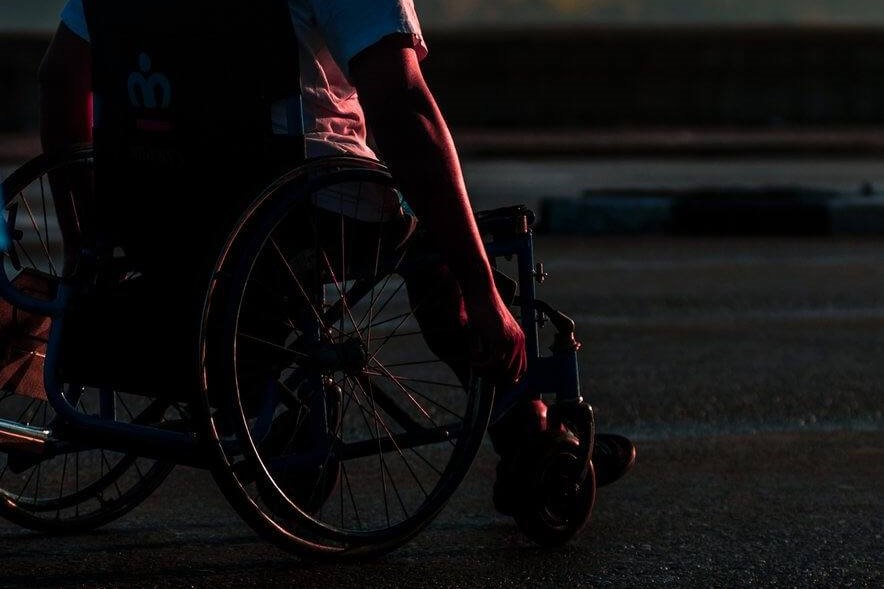
Photo by Ricardo IV Tamayo on UnsplashEditor’s Note: This page has been updated for accuracy and relevancy [cha 2.25.21]